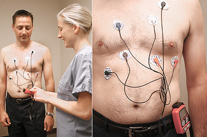
68 Harley Street London, W1G 7HE · Main Office
Also at Cromwell & Syon Bishops Wood · Multiple Locations
0203 9838 001 Call for Appointments
jessica@oneheartclinic.com Rapid Response to Enquiries
Expert London Cardiologist for your Heart Health
Cardiology has more imaging and diagnostic tools than almost any other specialty. Each test answers a different question — rhythm, structure, blood supply, muscle health. Here, Dr Sukhjinder Nijjer explains when each test is the right choice.
Dr Sukhjinder Nijjer BSc(Hons) MB ChB(Hons) PhD FRCP — Consultant Cardiologist with specialist interest in cardiac imaging
GMC: 6103417 · Harley Street · Cromwell Hospital · Syon Clinic · Bishops Wood Hospital · Last reviewed:
Dr Nijjer offers the full range of cardiac investigations — from a resting ECG to coronary angiography — at One Heart Clinic and partner hospitals. Most tests can be arranged at the same visit as your consultation.
Dr Nijjer performing a cardiac ultrasound at One Heart Clinic
| Test | What it shows | Time | Radiation | Best for |
|---|---|---|---|---|
| ECG (resting) | Rhythm, electrical activity, evidence of past heart attack | 5 min | None | First-line rhythm assessment |
| Echocardiogram | Chamber size, pumping function, valves, pressures | 20–40 min | None | Heart structure and function |
| Stress echocardiogram | Blood supply adequacy under exertion | 45–60 min | None | Ischaemia / angina assessment |
| CT coronary angiogram | Coronary artery anatomy and plaque | 10–15 min | Low | Ruling out coronary disease |
| Calcium score | Calcified plaque burden; 10-yr risk | 5–10 min | Very low | Risk stratification / statin decision |
| Cardiac MRI | Scar, inflammation, cardiomyopathy, accurate EF | 45–60 min | None | Heart muscle detail |
| Holter / patch monitor | Rhythm over 24 h–14 days | Worn continuously | None | Intermittent palpitations or syncope |
| Invasive angiogram | Coronary anatomy; allows same-session stenting | 20–40 min | Moderate | Confirmed or high-risk disease |
An ECG is a 10-second recording of the heart's electrical activity and excels at diagnosing rhythm problems, ongoing or previous heart attacks, thickened heart muscle, and certain inherited electrical conditions.
What it cannot do is show how the heart is moving or pumping, or whether the coronary arteries are narrowed at rest. A normal ECG between symptomatic episodes does not rule out arrhythmia or coronary disease — for those, you usually need a recording during symptoms (Holter or patch monitor) or an imaging test such as an echo, CT or MRI.
An echocardiogram is an ultrasound scan of the heart, showing the size and pumping function of each chamber, how well the heart valves are working, the thickness of the muscle, and the pressure in the pulmonary arteries.
Echocardiography uses no radiation, no needles and no contrast in its standard form, making it completely safe and repeatable. A standard scan takes 20 to 40 minutes. A more detailed variant — the transoesophageal echo — passes a probe down the throat for a closer look and is reserved for specific situations where the standard scan is limited.
"The echocardiogram is my most versatile tool. In a single 30-minute scan I can assess pumping function, valves, pressures and structure — all without any radiation or discomfort. It's often the first test I arrange, and it answers more questions than almost anything else in the clinic."
— Dr Sukhjinder Nijjer, Consultant Cardiologist
A standard exercise ECG records electrical activity while you walk on a treadmill, looking for electrical signs of inadequate blood supply during exertion. It is inexpensive and widely available but only modestly accurate — particularly in women.
A stress echocardiogram combines exercise (or a chemical that mimics it) with ultrasound imaging before, during and after the stress. The cardiologist looks for areas of heart muscle that move less well under load, which signals a meaningful blockage in the artery supplying that region. Stress echo is significantly more accurate and has become the preferred functional test in most UK practice.
In most patients with new chest pain, NICE recommends starting with a CT coronary angiogram — a non-invasive scan that gives a detailed look at the coronary arteries in about ten minutes. It is excellent at ruling out significant coronary disease and identifying who needs further investigation.
An invasive coronary angiogram is reserved for patients with definitive evidence of significant disease on CT or stress testing, those with high-risk symptoms such as unstable angina or a recent heart attack, or those in whom CT cannot be performed reliably. The big advantage of the invasive angiogram is that it can move directly to treatment — placing a stent in the same procedure if a significant blockage is confirmed.
The echocardiogram is probably my single most used diagnostic tool. In 20–30 minutes, I can see the heart contracting in real time, assess valve function, measure pressures and detect structural abnormalities that no blood test reveals. Patients are often surprised to learn more about their heart from a single echocardiogram than they have in decades.
Beyond standard imaging, I use iFR pressure wire assessment — technology I co-developed — to measure blood flow across coronary narrowings. This is the most precise way to determine whether a stent is actually needed, and it is now used in patients worldwide and recommended in international guidelines. For complex coronary anatomy I also use intracoronary imaging (IVUS and OCT) to guide treatment decisions with a level of precision that angiography alone cannot provide.
No preparation needed. Electrodes placed on chest, arms and legs. Completely painless. Results available immediately.
No preparation needed. Ultrasound gel applied to chest. Completely painless. Detailed report produced at the same visit.
No eating for 4 hours before. Beta-blocker may be given to slow the heart rate. Contrast dye injected. Results within 1–2 days.
Fast from midnight. Wrist or groin access under local anaesthetic. Stent placed same day if needed. Most patients go home same day.
A coronary calcium score is a quick, low-dose CT scan that measures calcified plaque in the coronary arteries and translates the result into a 10-year risk of heart events. It takes about ten minutes, requires no contrast and no needles.
It is most useful in middle-aged adults at intermediate cardiovascular risk where the decision about starting a statin is genuinely uncertain — a score of zero argues strongly against starting, while a high score (over 100) argues firmly for it. It is not generally recommended in low-risk under-40s or in patients with established heart disease, where treatment is already indicated regardless.
"A calcium score is one of my favourite tools for the patient who isn't sure whether they need a statin. The result is often decisive — a score of zero gives genuine reassurance, while a high score makes the treatment conversation straightforward. It is one of the few tests where a single number genuinely changes a clinical decision."
— Dr Sukhjinder Nijjer, Consultant Cardiologist
Cardiac MRI is the most detailed imaging test for the heart muscle and uses no radiation. Its unique strengths are detecting scar tissue from previous heart attacks, identifying inflammation such as myocarditis, characterising cardiomyopathies, and measuring ejection fraction with the highest accuracy.
It is particularly valuable when an echocardiogram is inconclusive, when scar or inflammation is suspected, or when an inherited heart condition is being assessed. The scan takes 45 to 60 minutes, requires you to lie still and follow breath-hold instructions, and usually involves an injection of contrast.
Modern CT coronary angiograms use very low radiation doses — typically equivalent to 6 to 18 months of natural background radiation — and protocols continue to improve year on year. For most patients, the diagnostic benefit comfortably outweighs the small theoretical risk.
In younger patients, women of childbearing age and patients needing multiple scans, the radiation question is weighed more carefully. Cardiac MRI is often used instead when radiation avoidance is a priority, although the choice ultimately depends on the clinical question being asked.
Avoid caffeine for at least 12 hours before the test and a heavy meal for 3 hours. Wear comfortable clothes and shoes suitable for walking. Some medications, particularly beta-blockers, may need to be paused on the morning of the test — your cardiologist will advise specifically.
Bring a list of your medications. The test itself takes 30 to 60 minutes including preparation and recovery; the exercise portion is usually 6 to 15 minutes. Most people are back to normal activity immediately afterwards.
An invasive coronary angiogram is performed under local anaesthetic with mild sedation, almost always via the wrist (radial approach). A thin tube is guided up to the heart and X-ray dye is injected into the coronary arteries so the cardiologist can see narrowings or blockages in real time.
The procedure takes 20 to 40 minutes and is done as a day case. If a significant narrowing is found, a stent can often be placed in the same sitting. You can usually go home within a few hours, and most patients are back to normal activity within a couple of days — slightly longer if a stent has been placed.
"Most patients are pleasantly surprised by how straightforward an angiogram is. Via the wrist, there is very little discomfort and you're usually back to normal within a day or two. The information it provides — and the ability to treat at the same time — can be genuinely life-changing."
— Dr Sukhjinder Nijjer, Consultant Interventional Cardiologist
In private practice, most cardiac test results — ECG, echocardiogram, stress echocardiogram — are reported on the day of the test, with a formal letter sent within 24 to 48 hours. CT coronary angiograms and cardiac MRIs typically take 2 to 5 working days for the full report.
If an urgent result is identified during the scan — a critical narrowing, for example — you will be told immediately rather than waiting for the formal report. Time-critical findings always trigger same-day contact and onward planning.
The right test depends on your age, risk factors and the character of the pain. For most adults with new exertional chest pain, CT coronary angiogram is first-line under NICE guidance because it can both rule in and rule out coronary disease quickly and non-invasively.
If your pain is unlikely to be cardiac but further reassurance is wanted, an echocardiogram and exercise ECG may be sufficient. For patients with known coronary disease and a question of new ischaemia, a stress echocardiogram or cardiac MRI is usually preferred. The choice should always be discussed in a consultation — there is no one-size-fits-all answer.
No — a normal ECG between symptomatic episodes is reassuring but does not rule out coronary disease, intermittent arrhythmia, valve disease or cardiomyopathy. The ECG is a snapshot, not a film.
Many patients with significant coronary narrowings have completely normal resting ECGs. Many patients with palpitations have normal ECGs between episodes. If symptoms persist despite a normal ECG, more targeted testing — imaging, ambulatory rhythm monitoring or stress testing — is the appropriate next step.
Same-week access to ECG, echocardiography, stress echo, CT angiography and cardiac MRI across Dr Nijjer's London clinics, with results discussed in person.
Book a consultationConsultant Cardiologist with specialist interest in cardiac imaging and interventional cardiology. Trained at Hammersmith, Charing Cross, St Mary's, Royal Brompton and Harefield hospitals. PhD from Imperial College London (MRC-funded). Over 150 peer-reviewed publications. Lead for the Cardiac Catheter Labs, Hammersmith Hospital.
GMC: 6103417 · Harley Street · Cromwell Hospital · Syon Clinic · Bishops Wood Hospital
This page provides general information for educational purposes and is not a substitute for personalised medical advice. If you are concerned about symptoms, contact a healthcare professional. In a medical emergency, call 999.